-
Analysis Imaging
Diagnostic imaging is a sophisticated clinical procedure that helps detect ailment. It is also used to keep an eye on a client’s health and wellness and examine exactly how they respond to treatment. Get more enlightened about diagnostic imaging department on this blog.
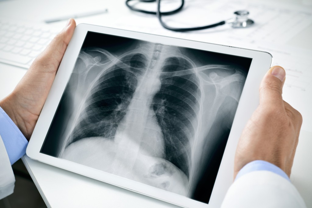
This type of clinical imaging utilizes electromagnetic radiation, as well as particular various other modern technologies, to develop images of the within the body. A variety of diagnostic imaging techniques are made use of, consisting of X-rays, CT scans, ultrasound and magnetic vibration imaging. Each has its very own advantages as well as negative aspects, and also is ideal matched for particular clinical problems. X-rays supply an aesthetic assessment of the bones and cells. These pictures are typically helpful in diagnosing cracks, bone injuries, as well as osteoporosis. X-rays are painless and fast. They are usually extracted from a selection of angles, and can be taped onto CDs. An additional diagnostic imaging strategy is arthrogram. Utilizing x-rays, fluoroscopy, and various other sorts of imaging, medical professionals can view the joints.
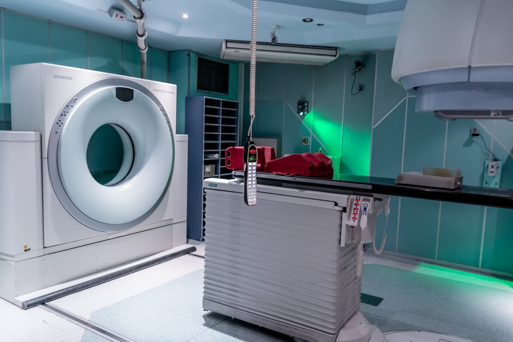
Arthrograms are valuable in detecting joint troubles. The joint is coated with iodine, permitting the radiologist to identify abnormalities. Shots of contrast dye emphasize areas that are not noticeable on x-rays. When integrated with various other analysis imaging methods, such as ultrasound, arthrograms are an effective way to detect a range of joint problems. CT scans are an advanced form of X-rays. These electronic tomography scans combine X-rays and also computer system scans to generate a cross-sectional photo of the inner frameworks of the body. A CT scan can be beneficial in identifying heart disease, lung condition, and various other conditions. The images can be used to aid medical professionals choose if surgical treatment is essential, as well as can assist the physician during biopsy treatments. Click here to get more details about analysis imaging.
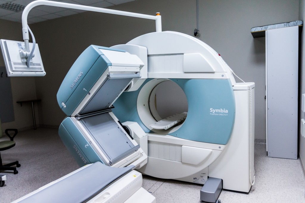
MRI (magnetic vibration imaging) is an alternative to X-rays. An MRI is extra accurate than a regular x-ray. MRIs are a closed-design imaging equipment that provides comprehensive photos of the human body. MRIs are commonly made use of to check out individuals with internal injuries, such as broken bones. Diagnostic imaging is a major part of health care today, and also its prevalent use has added to enhanced patient care. With exact images, clients can make even more informed clinical choices. Also, diagnostic imaging modern technology can be made use of to recognize illness at an earlier stage, leading to reduced expenses and faster therapies. Ultrasound is a preferred type of diagnostic imaging. This medical strategy is frequently made use of in obstetrics to look for pregnancy, and also for fetal monitoring.
For the most part, this technique is painless, yet there are some safety measures to keep in mind. If you have deodorant or lotion on your underarms, this can disrupt the imaging process. CT scans are additionally valuable for identifying heart as well as lung conditions, in addition to inner blood loss. They can likewise be useful in identifying the place of a lump. Similarly, bone density scans are a typical medical imaging technique. Bone thickness is reduced with age. X-rays and also ultrasound are good ways to review bone stamina, while bone thickness scans can be done by a handheld gadget or a health center bed. Radio-opaque comparison media can be made use of to visualize the intestine and belly, however can also be used to establish the existence of abscess, along with certain types of colon cancer cells. For more details about this subject, click here: https://en.wikipedia.org/wiki/Medical_imaging.
-
Diagnostic Imaging

Diagnostic imaging is an essential tool to assist medical professionals in examining as well as establishing the cause of disease, injury or illness. This team of techniques consists of x-rays, ultrasound, computed tomography (CT) scans and also magnetic vibration imaging (MRI). These approaches make it possible for doctors to see inside of the person, enabling them to evaluate the framework and also function of organs, tissues and also bones.
The stayton surgical center also offer high-resolution pictures that allow for even more exact diagnosis. Imaging is necessary to determine the existence of a growth, to determine the place of a heart clot or to look for fractures. It additionally assists in evaluating the action of a treatment. While most of the imaging tests are non-invasive, some might require the client to stay still for extended periods of time. If the doctor requests numerous views or zooms, the test might take longer. X-rays are one of the most common sort of analysis imaging, although various other techniques exist. X-rays are a non-invasive treatment that includes a high-energy beam of light given off from an x-ray equipment.
The rays pass through the body and create a black-and-white photo of the bones as well as lungs. A CT check is an advanced X-ray that makes use of a number of X-rays extracted from different angles to develop a cross-sectional picture of the body. Depending on the circumstance, the radiologist might make a decision that a CT check is a lot more efficient than a basic X-ray. Ultrasound and radio-opaque contrast media are also typical forms of diagnostic imaging. During pregnancy, ultrasound is typically used to keep track of the unborn child. Additionally, using radio-opaque comparison media allows physicians to imagine the interior structures of the belly and also colon. A number of researches have shown that making use of these devices has boosted substantially over the last a number of years. MRIs are also popular, specifically for people with inner injuries. Click here for more details concerning diagnostic imaging.
MRIs supply an even more detailed photo than normal x-rays, and are especially beneficial for finding bone fractures and other inner trauma. They can additionally be used to aid detect capillary issues as well as cancer. In a shut MRI, a patient lies down as well as the source of a high-energy x-ray beam rotates around the individual’s body, creating an image. Other common imaging treatments include arthrograms, which are pictures of the joints. They can be made use of to detect joint conditions. One more form of imaging is MRA scans, which are utilized to spot blood vessel concerns. Making use of a mix of x-rays and also computer software, cross-sectional photos are generated. Analysis imaging has actually been a tremendous source for people and also medical professionals.
Nonetheless, it has not been an affordable choice for many low-income countries, which do not have educated medical care workers and do not have the sources to buy imaging tools. There are numerous reasons for this. One factor is the high cost of imaging, which might be as high as $100 billion yearly. One more factor is the health and wellness risks connected with radiation exposure. Medical imaging is essential at every major level of healthcare. The use of analysis imaging technology has improved person results as well as decreased the requirement for unnecessary exploratory procedures. Nonetheless, information to measure the benefits of imaging are restricted. As a result, it is necessary that evidence-based guidelines balance the risks as well as benefits of imaging. For better understanding of this topic, please click here: https://en.wikipedia.org/wiki/Radiology.
-
The Benefits as well as Dangers of Diagnostic Imaging

Diagnostic imaging describes medical strategies which make it possible for medical professionals to look inside of a person’s body and also to determine what is triggering a health problem. They can additionally figure out the training course of an illness. The results of analysis imaging can aid physicians to make an enlightened choice regarding therapy as well as safety nets. It is likewise vital to record a client’s reaction to treatment. Clinical imaging has actually come to be a central component of healthcare in the 21st century. This is due to its advantages, that include earlier medical diagnosis, better treatment options, as well as a lot more precise medical diagnoses. Get the benefits of diagnostic imaging here: https://santiamhospital.org/services/surgical-center/.
Several kinds of imaging procedures are non-invasive and also painless. Nevertheless, some treatments include small amounts of radiation. There are additionally wellness dangers related to radiation exposure. Those dangers consist of cancer and various other conditions. Utilizing evidence-based guidelines, healthcare providers need to stabilize the benefits of imaging with the risks of its usage. While medical imaging has been used in a range of ways, one of the most commonly used kinds are x-rays, CT checks, and also MRIs. These treatments generate photos of the inside of the body, consisting of bones and organs. On top of that, they can spot fractures, cancer, embolism, as well as various other wellness conditions.
A number of these tests are non-invasive as well as take just a few minutes to complete. X-rays are a type of diagnostic imaging that includes the use of high-energy light beams to develop images of the inside of an individual’s body. Some individuals might experience pain throughout the process, yet most examinations are painless. If you have any type of issues about the procedure, speak with your doctor. X-rays are largely utilized for diagnosing injuries, such as a fractured bone. Numerous elements affect the amount of radiation that a person is revealed to throughout the test. A low-energy x-ray can discover osteoporosis. On the various other hand, an extra powerful x-ray can show indicators of a cancer growth. Click here to get more info about diagnostic imaging.
CT and MRI are more advanced analysis imaging approaches that enable physicians to see inside a person’s body. During these treatments, a person rests on a table while the device takes a series of images. Each photo is then integrated with others to develop a two-dimensional photo. CT and also MRI images are frequently beneficial in detecting as well as treating heart and lung illness. An MRI uses electromagnetic fields to produce a photo of a person’s mind, while a CT scan can be used to check out other body organs. X-rays are the most common diagnostic imaging strategy. They are painless and can be quickly executed on people of all ages. X-rays can be extracted from a range of angles, and are generally finished in simply ten minutes.
The majority of physicians will certainly create the x-ray photo to a CD. Ultrasound, which creates images of body organs utilizing acoustic waves, is an additional typical type of diagnostic imaging. While pregnant, it is utilized to check the growth of a fetus. Similarly, arthrograms are made use of to analyze joints. While clinical imaging is an integral component of medical care today, there is restricted information on the benefits of the innovation. Nevertheless, the Globe Health Organization (THAT) and its partners work to advertise client safety and security by collaborating with manufacturers and also supplying training programs. This post: https://en.wikipedia.org/wiki/Magnetic_resonance_imaging, will help you understand the topic even better.
-
Diagnostic Imaging
Analysis imaging entails a number of approaches for seeing inside the body. These methods are developed to aid physicians recognize the reason for an ailment or injury and also monitor treatment. It is likewise used to validate a medical diagnosis. X-rays are one of the most commonly utilized sort of diagnostic imaging. An x-ray equipment generates a high-energy light beam, which passes through the body and also produces a photo. This picture is after that refined to produce a cross-sectional sight of the interior organs. X-rays are likewise made use of to identify busted bones as well as to check out joints. MRI is a more advanced kind of diagnostic imaging. Click here: https://santiamhospital.org/services/diagnostic-imaging/, to read more about analysis imaging.
Unlike X-rays, which make use of electromagnetic waves to produce an image, MRI utilizes radio waves, which develop a detailed fixed picture on a movie. MRI has boosted recently. Making use of an extra effective electromagnetic field, MRI provides an extra exact sight of the body. Ultrasound is an additional usual diagnostic imaging method. Powered by sound waves, ultrasound can envision the inside of the body. While pregnant, ultrasound is typically used to monitor the advancement of the unborn child. In the biliary tract, ultrasound is specifically sensitive. Various other kinds of diagnostic imaging consist of computed tomography, or pet cat scans.
CAT scans are made use of to detect embolism, tumors, and also fractures. They are additionally valuable throughout a procedure to lead the doctor. CT scans are likewise an innovative form of X-ray. CT scans produce cross-sectional images of the body, and also they can help medical professionals throughout surgical treatments as well as throughout radiation therapy. Since they are a non-invasive technique, CT scans are pain-free. Nonetheless, they are not as precise as X-rays. The rate of CTs among Medicare fee-for-service enrollees was 550 per 1000 enrollees in 2006. Nuclear medication is one more diagnostic imaging modality. Numerous radioactive isotopes are injected into cells to give pictures. Although radiation is entailed, it is connected with a tiny danger. Read more about this services on this blog.
A CT scan is especially reliable in instances of inner trauma, because it can supply a clear sight of the entire body. Advancements in analysis imaging have actually boosted the quality of person care. Along with an extra exact medical diagnosis, the modern technology can aid individuals prevent unnecessary exploratory procedures. As a result, the use of innovative diagnostic imaging has actually enhanced dramatically. Over the past fifteen years, 30.9 million imaging assessments were carried out by Medicare enrollees. Most of these examinations are executed by a radiologist. Imaging prices vary between health and wellness systems and also areas.
Distinctions in health condition and age might account for these distinctions. Health plans as well as HMOs may have reduced imaging rates. Also, several low-income nations can not afford to acquire devices for diagnostic imaging. In addition, defensive medical methods have likely contributed to the increase being used. The that as well as various other partners emphasize person safety and work together with producers to develop technical solutions. Evidence-based standards ought to be established to balance the benefits of imaging with the dangers to the patient. A number of these methods are non-invasive, decreasing the quantity of ionizing radiation that the patient gets. Get a general overview of the topic here: https://en.wikipedia.org/wiki/Radiography.
-
Analysis Imaging
Diagnostic imaging is a term that refers to numerous different non-invasive methods for seeing inside the human body. These examinations can help doctors detect and also monitor the efficiency of treatments. They can additionally detect diseases ahead of signs and symptoms developing. However, before going through any imaging procedure, people should reveal their medical history, including any type of drugs, and also allow their medical professionals know of any kind of other clinical problems they may have. One of the simplest types of analysis imaging is X-rays. Click to read more about analysis imaging.
This test is a pain-free way to picture bones. It is frequently utilized to analyze fractures. A CT check is a less intrusive type of X-ray that produces in-depth cross-sectional photos of the body. A magnetic resonance imaging (MRI) scan is a more advanced approach that uses extremely powerful magnets and also computer system technology to create a more detailed picture of the inside of the body. Analysis imaging can be done in both healthcare facilities and clinics. The treatment is usually finished in a matter of mins. Various devices is used to produce the pictures. A few of the equipment consists of x-ray machines, a motorized X-ray source, as well as a computer system. In addition to the equipment, a radiologist, or a medical professional trained in analyzing x-rays, is involved. An additional diagnostic imaging device is an ultrasound tool. Read more about diagnostic imaging here: https://santiamhospital.org/.
This technology is powered by high-frequency acoustic waves and permits the physician to see the structure of the intestines, belly, as well as various other components of the body. While pregnant, ultrasound is very important to keep track of the fetus. Ultrasound can likewise be utilized to discover breast cancer cells. Analysis imaging is an essential part of validating a disease’s training course and documenting its feedback to treatment. Although radiation is associated with a little threat, there is an overwhelming clinical advantage. For instance, a CT scan can discover abnormalities in the bones, heart, and blood vessels. Radiation exposure is connected with a reduced rate of cancer cells, yet there are concerns regarding malpractice.
There are many various other imaging treatments that are not as popular. Examples include MRA and animal scans. Magnetic resonance imaging can be made use of to check out malformations of blood vessels. Lots of low-income countries do not have the resources to purchase and also maintain these kinds of equipment, but they are indispensable in some locations. Advanced analysis imaging has changed substantially throughout the years. Earlier detection of a problem is valuable, as is lower healthcare prices. While the majority of these procedures are painless, some require a patient to stay still for a period of time. For that reason, it is important to talk about the possible dangers and benefits of the process.
When the benefits outweigh the risks, evidence-based guidelines can be created to make informed choices concerning treatment. Among Medicare fee-for-service enrollees, X-rays and CTs were the most commonly utilized analysis imaging treatments in 2006. A number of other modalities were likewise made use of, such as radiography as well as angiography/fluoroscopy. The number of tests carried out annually varied dramatically. Diagnostic imaging prices raised quickly during the early 2000s. These boosts were driven by both consumer need and defensive medical practices. HMO use imaging services additionally raised dramatically. By 2006, the price for MRIs among HMO enrollees aged 65 as well as older was 123 per 1000 enrollees. Education is a never ending process, so continue reading here: https://en.wikipedia.org/wiki/Medical_diagnosis.
-
Subscribe
Subscribed
Already have a WordPress.com account? Log in now.
